80-year-old Russian woman survived with needle in brain after parents’ failed World War II infanticide

Needle-in-Brain

Important Takeaways:

  • An 80-year-old Russian woman has lived her entire life with a needle measuring over an inch in length lodged in her brain, stunned doctors have revealed.
  • Medics on the remote Russian island of Sakhalin in the Pacific Ocean have concluded that the needle likely was proof of her parents’ botched attempt to kill her as a newborn during World War II.
  • “Such incidents were not uncommon during the years of starvation: a thin needle would be inserted into a newborn’s fontanel to damage the brain,”
  • “The fontanel quickly closed up, covering up evidence of the crime, and the baby died.”
  • In the case of the elderly patient, the 1.2-inch-long needle pierced her left parietal lobe, but that “did not lead to the intended effect,”
  • The baby girl survived the attempted infanticide — and in the coming years and decades she had never complained of headaches due to the needle nestled in her brain

Read the original article by clicking here.

Brain and spine implants allow a paralyzed man to walk again

Daniel 12:4 But you, Daniel, roll up and seal the words of the scroll until the time of the end. Many will go here and there to increase knowledge.”

Important Takeaways:

  • A Paralyzed Man Can Walk Naturally Again With Brain and Spine Implants
  • Gert-Jan Oskam was living in China in 2011 when he was in a motorcycle accident that left him paralyzed from the hips down. Now, with a combination of devices, scientists have given him control over his lower body again.
  • “For 12 years I’ve been trying to get back my feet,” Mr. Oskam said in a press briefing on Tuesday. “Now I have learned how to walk normal, natural.”
  • In a study published on Wednesday in the journal Nature, researchers in Switzerland described implants that provided a “digital bridge” between Mr. Oskam’s brain and his spinal cord, bypassing injured sections. The discovery allowed Mr. Oskam, 40, to stand, walk and ascend a steep ramp with only the assistance of a walker. More than a year after the implant was inserted, he has retained these abilities and has actually showed signs of neurological recovery, walking with crutches even when the implant was switched off.
  • “We’ve captured the thoughts of Gert-Jan, and translated these thoughts into a stimulation of the spinal cord to re-establish voluntary movement,” Grégoire Courtine, a spinal cord specialist at the Swiss Federal Institute of Technology, Lausanne, who helped lead the research, said at the press briefing.
  • Oskam had undergone stimulation procedures in previous years, and had even regained some ability to walk, but eventually his improvement plateaued. At the press briefing, Mr. Oskam said that these stimulation technologies had left him feeling that there was something foreign about the locomotion, an alien distance between his mind and body.
  • The new interface changed this, he said: “The stimulation before was controlling me, and now I’m controlling the stimulation.”

Read the original article by clicking here

German scientists create see-through human organs

An employee works with stem cells at Dr. Ali Ertuerk's laboratory in Munich, Germany April 23, 2019. Mr. Ertuerk and his team developed DISCO transparency technology which is used by scientists from diverse biomedical research fields to generate high resolution views of intact rodent organs and bodies, a milestone on the way to generate 3D-bioprinted human organs. Picture taken April 23, 2019. REUTERS/Michael Dalder

By Ayhan Uyanik

MUNICH (Reuters) – Researchers in Germany have created transparent human organs using a new technology that could pave the way to print three-dimensional body parts such as kidneys for transplants.

A transparent mouse is pictured at the laboratory of Dr. Ali Ertuerk, Group Leader at the Institute for Stroke and Dementia Research (ISD) at the Ludwig Maximillian's University, in Munich, Germany April 23, 2019. Mr. Ertuerk and his team developed DISCO transparency technology which is used by scientists from diverse biomedical research fields to generate high resolution views of intact rodent organs and bodies, a milestone on the way to generate 3D-bioprinted human organs. REUTERS/Michael Dalder

A transparent mouse is pictured at the laboratory of Dr. Ali Ertuerk, Group Leader at the Institute for Stroke and Dementia Research (ISD) at the Ludwig Maximillian’s University, in Munich, Germany April 23, 2019. Mr. Ertuerk and his team developed DISCO transparency technology which is used by scientists from diverse biomedical research fields to generate high resolution views of intact rodent organs and bodies, a milestone on the way to generate 3D-bioprinted human organs. REUTERS/Michael Dalder

Scientists led by Ali Erturk at Ludwig Maximilians University in Munich have developed a technique that uses a solvent to make organs such as the brain and kidneys transparent.

The organ is then scanned by lasers in a microscope that allows researchers to capture the entire structure, including the blood vessels and every single cell in its specific location.

Using this blueprint, researchers print out the scaffold of the organ. They then load the 3D printer with stem cells which act as “ink” and are injected into the correct position making the organ functional.

While 3D printing is already used widely to produce spare parts for industry, Erturk said the development marks a step forward for 3D printing in the medical field.

Until now 3D-printed organs lacked detailed cellular structures because they were based on images from computer tomography or MRI machines, he said.

“We can see where every single cell is located in transparent human organs. And then we can actually replicate exactly the same, using 3D bioprinting technology to make a real functional organ,” he said.

A transparent human brain is shown by Dr. Ali Ertuerk at his laboratory in Munich, Germany April 23, 2019. Mr. Ertuerk and his team developed DISCO transparency technology which is used by scientists from diverse biomedical research fields to generate high resolution views of intact rodent organs and bodies, a milestone on the way to generate 3D-bioprinted human organs. Picture taken April 23, 2019. REUTERS/Michael Dalder

A transparent human brain is shown by Dr. Ali Ertuerk at his laboratory in Munich, Germany April 23, 2019. Mr. Ertuerk and his team developed DISCO transparency technology which is used by scientists from diverse biomedical research fields to generate high resolution views of intact rodent organs and bodies, a milestone on the way to generate 3D-bioprinted human organs. Picture taken April 23, 2019. REUTERS/Michael Dalder

“Therefore, I believe we are much closer to a real human organ for the first time now.”

Erturk’s team plan to start by creating a bioprinted pancreas over the next 2-3 years and also hope to develop a kidney within 5-6 years.

The researchers will first test to see whether animals can survive with the bioprinted organs and could start clinical trials within 5-10 years, he said.

(Reporting by Ayhan Uyanik; Writing by Caroline Copley; Editing by Susan Fenton)

Study in mice shows Zika virus also attacks adult brain cells

Aedes aegypti mosquitoes are seen inside Oxitec laboratory in Campinas, Brazil,

By Julie Steenhuysen

CHICAGO (Reuters) – U.S. researchers have found that Zika can attack special populations of brain cells in adult mice in the part of the brain involved in learning and memory, raising new questions about how the virus may be impacting millions of adults who have been infected with the virus.

The findings, published on Thursday in the journal “Cell Stem Cell,” are the first to look at whether Zika can attack the same kinds of cells in adult mice that they do in fetal mice.

Experts cautioned that the findings are preliminary and may not have any correlation to how Zika impacts human brain function, but they suggest the need for follow-up research.

“This is one potential consequence we need to look at,” said Dr. Joseph Gleeson, an expert in pediatric brain disease at The Rockefeller University in New York, who led the study.

Zika has already been shown to attack fetal brain cells known as neural progenitor cells – a type of stem cell that gives rise to various kinds of brain cells. The death of these cells is what disrupts brain development and leads to the severe birth defects seen in babies whose mothers were infected with Zika during pregnancy.

U.S. health officials have concluded that Zika infections in pregnant women can cause microcephaly, a birth defect marked by small head size that can lead to severe developmental problems in babies.

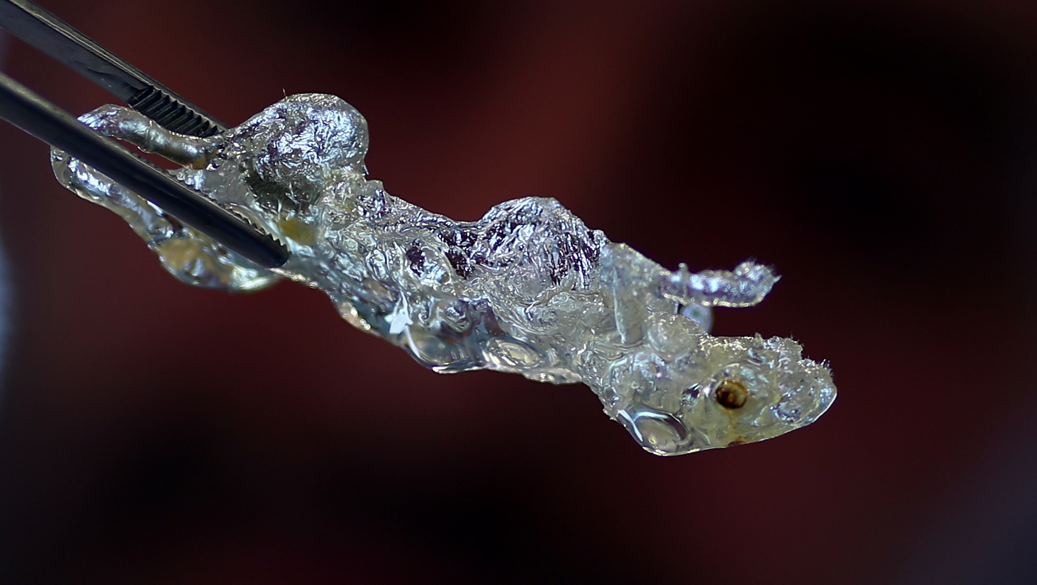
The connection between Zika and microcephaly first came to light last fall in Brazil, which has now confirmed more than 1,600 cases of microcephaly that it considers to be related to Zika infections in the mothers.

Fetal brains are chock full of neural progenitor cells, which are responsible for making cells that form key brain structures. Adults, whose brains are fully formed, have far fewer and there are some pockets remaining – including in the hippocampus, a part of the brain involved in memory and learning.

Gleeson wanted to see if Zika could attack these cells in adult mice. To find out, his team injected the virus into lab mice and examined their brains for Zika infection.

In the hippocampus, Gleeson said “it lit up like a Christmas tree and wiped out the stem cell population.”

“Based on our findings, getting infected with Zika as an adult may not be as innocuous as people think,” he said.

There have already been signs that Zika affects adult nerve cells. Several teams have published papers showing that in some patients, Zika can cause serious brain and spinal cord infections – including encephalitis, meningitis and myelitis – in people exposed to Zika.

In rare cases, Zika has also been linked with Guillain-Barre Syndrome, a post-infectious autoimmune disorder that can cause temporary paralysis in adults.

“It’s really unclear if this translates to human Zika infections,” said Dr. Daniel Pastula, a neurologist and medical epidemiologist at the University of Colorado Denver.

But if it does, it is not clear whether the effect is temporary or lasting.

“Detailed neurological studies are needed in infected humans to describe the effects of Zika virus infection on the brains of adults,” said Dr. Anna Checkley of the Hospital for Tropical Diseases, part of University College London Hospitals.

Gleeson said the study needs to be replicated by other scientists, and he wants to test other strains of Zika on adult mice at other times during an infection to see if those viruses have the same effect.

Since 2015, 66 countries and territories have reported evidence of vector-borne Zika virus transmission, according to the World Health Organization.

Earlier this month, researchers at Notre Dame University estimated as many as 93 million people across Latin America and the Caribbean could become infected with Zika in the current outbreak.

(Reporting by Julie Steenhuysen, editing by G Crosse)